Research
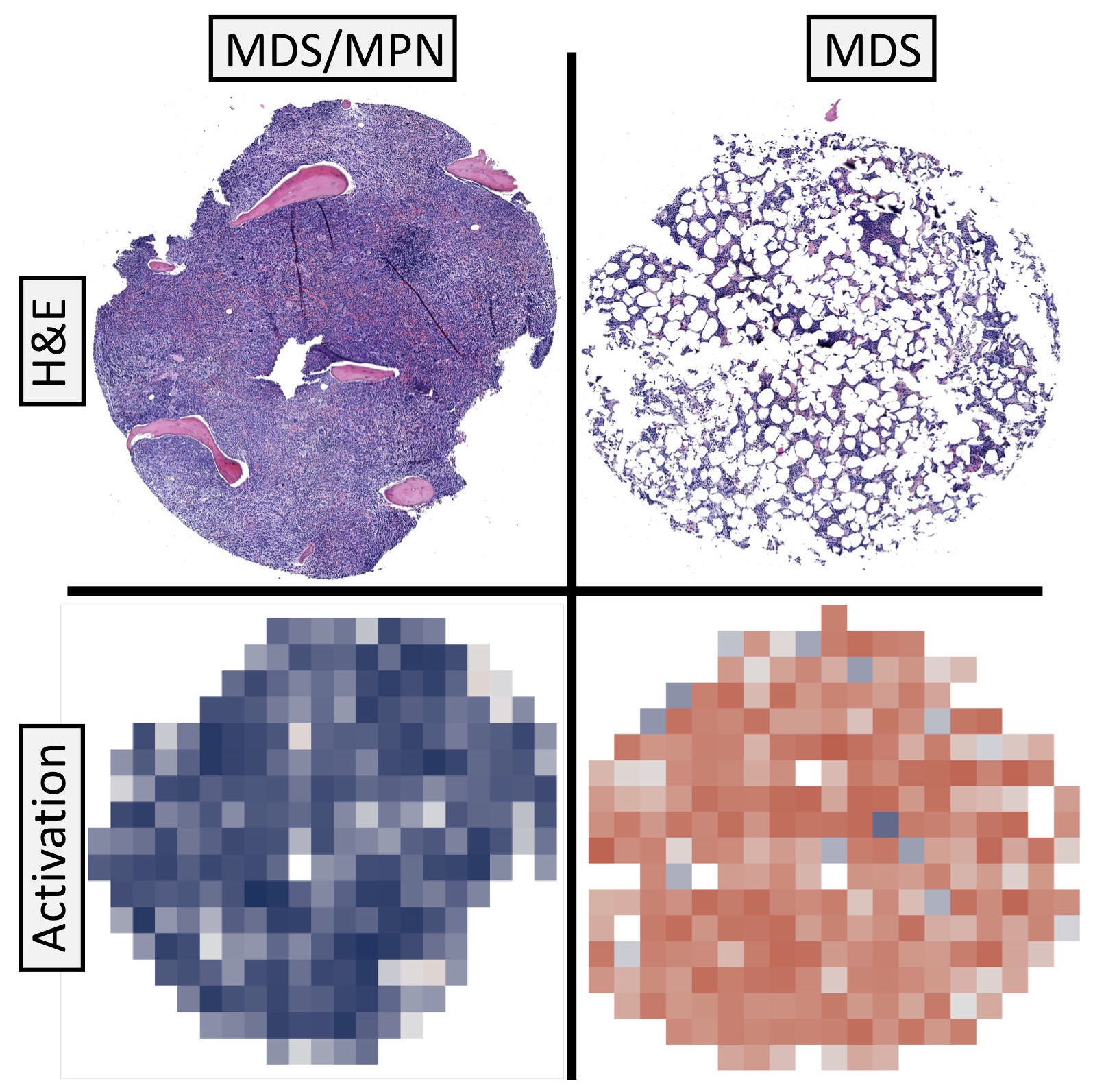
In hematology, there is an acute unmet need for automated machine vision to speed up routine diagnostics due to persistant shortage of trained laboratory physicians and increasing number of analyzed samples per year. Our recent results suggest that peripheral blood and bone marrow samples have an unexplored potential in disease subclassification, with direct possibilities of translation to patient prognostication (Brück et al, 2021).
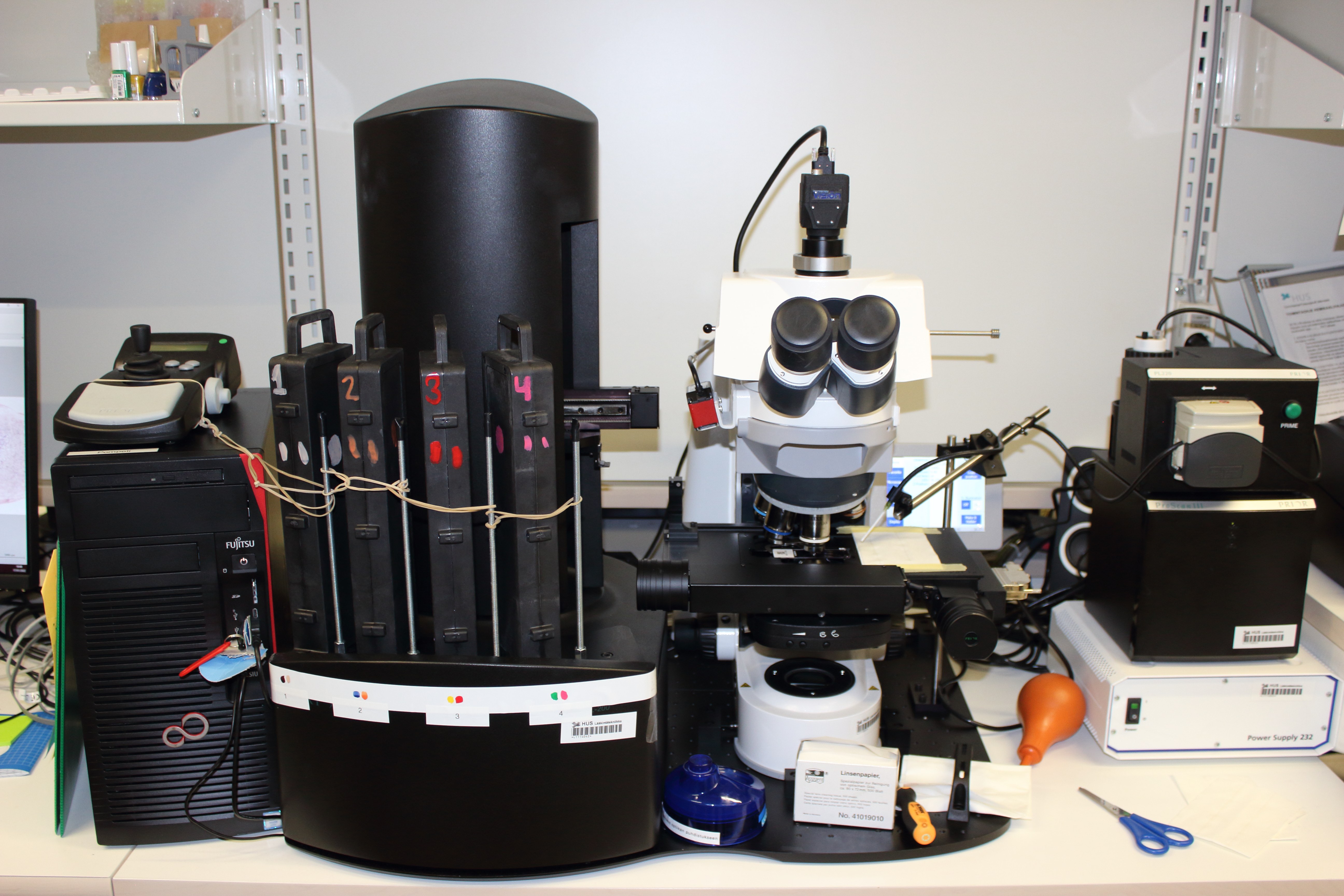
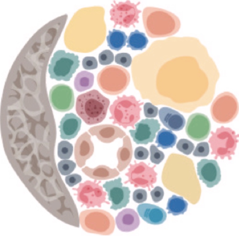
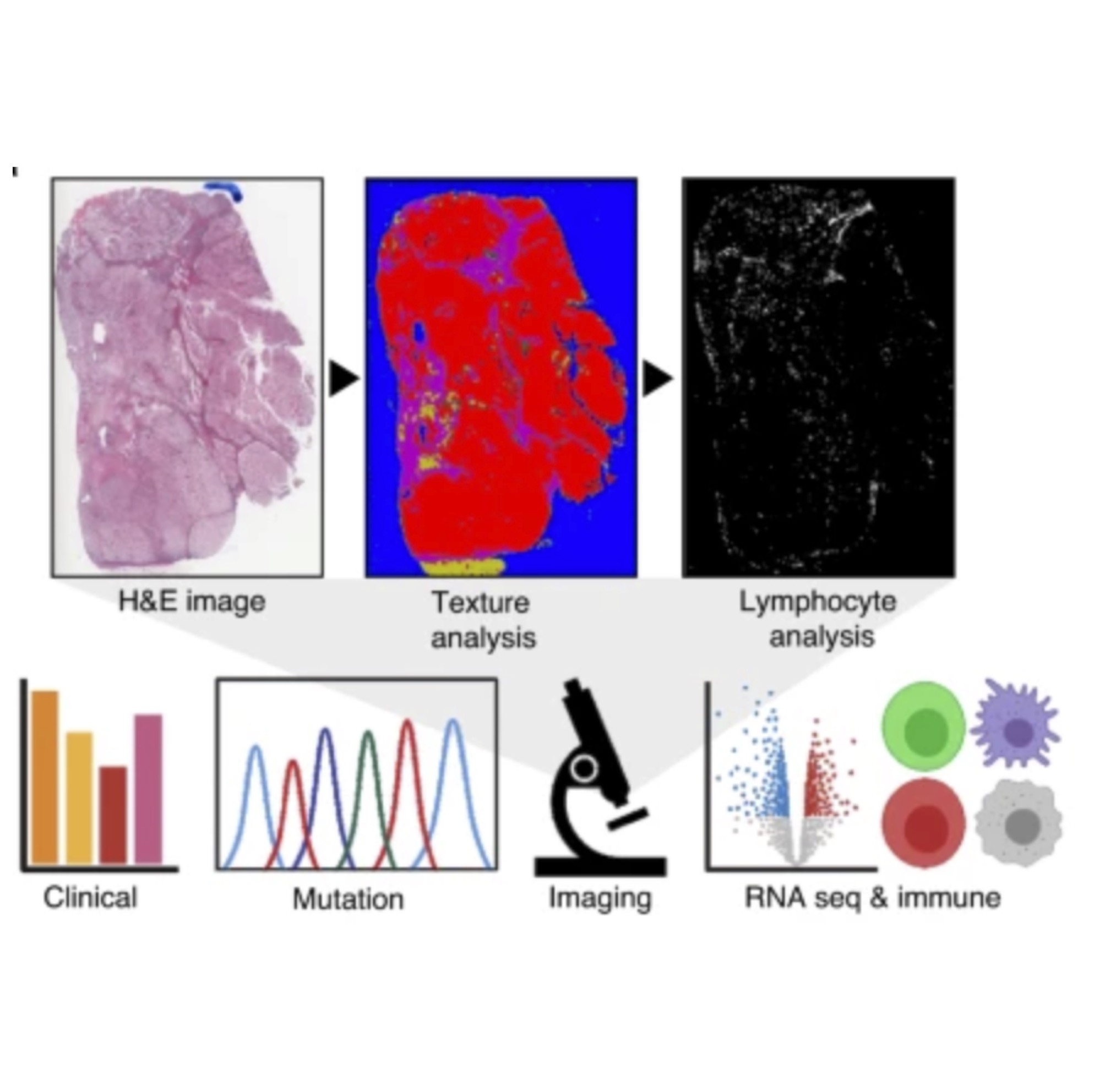
We have set up a digital slide scanner in the Helsinki University Hospital laboratory (HUSLAB). We have digitized standard cytomorphological samples into an extensive digital image archive. The second aim of the project is to develop image analysis algorithms to decode cell morphologies present in the hematocytological samples. Our software will replicate cytopathological examination of bone marrow and blood samples by detecting and classifying cells into 17 different types, inform of signs of dysplasia, and notify on the sample’s general composition. In addition, the software can identify novel biomarkers of treatment.
Computer-assisted diagnostics will reduce time required for slide examination, provide deeper information on risk and treatment stratification, and help us to better understand the biology of various disease phenotypes. The extensive image archive will serve both in terms of quality control of bone marrow products, standardize the training of specializing physicians, and improve future collaboration with hospitals, research groups, and companies. In summary, we will build a globally unique image dataset of hematological patients with the ultimate aim to develop and integrate severely needed algorithms into clinical practice.


